
Now Reading: Laminae As Potential Biosignatures
-
01
Laminae As Potential Biosignatures
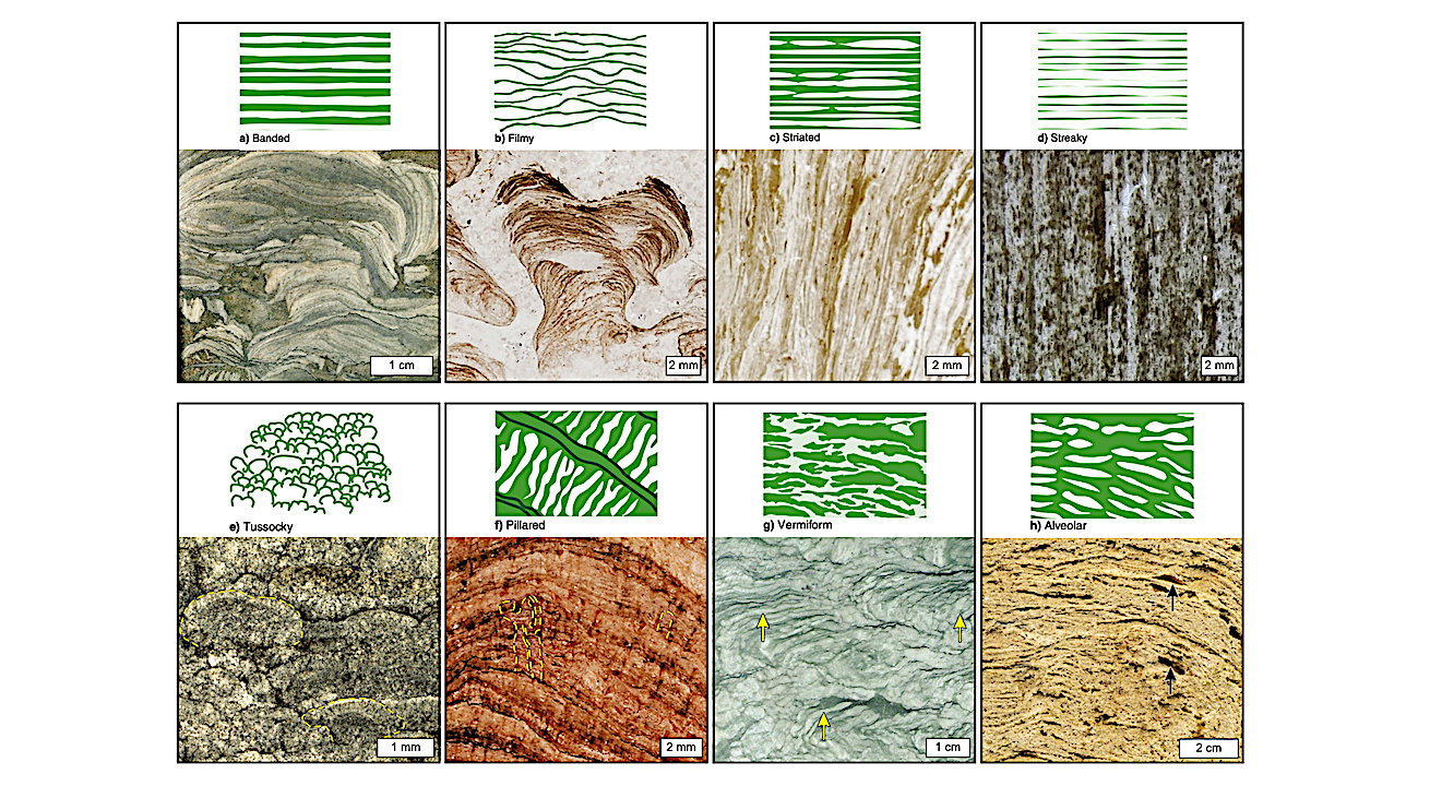

Figure 5.) Stromatolite lamination architectural types and associated examples, reproduced from Grey, K and Awramik, S.M. 2020, Handbook for the Study and Description of Microbialites (Geological Survey of Western Australia, Bulletin 147, licensed under Creative Commons Attribution 4.0 International Licence). A. Banded lamination type; example is from Paleoproterozoic-aged Peedawarra Flats, Mount Phillips, Western Australia (polished face, sample ID: GSWA F9908–46036; photo by K. Grey). B. Filmy lamination type; example is from Paleoproterozoic-aged Nabberu Basin, Western Australia (thick section, sample ID: GSWA F12365–46333; photo by S.M. Awramik and K. Grey). C. Striated lamination type; example is from Paleoproterozoic-aged Turee Creek, Western Australia (thick section, sample ID: GSWA F53603–84727; photo by S.M. Awramik and K. Grey). D. Streaky lamination type; example is Mesoproterozoic-aged from Pingandy Creek, Mount Egerton, Western Australia (thick section, sample ID: GSWA F9932–46009; photo by M. Ang). E. Tussocky lamination type (outlined); example is Paleoproterozoic-aged from near Lake Wells Homestead, Throssell, Western Australia (thick section, sample ID: GSWA F52286–193359; photo by K. Grey). F. Pillared lamination type (outlined); example is Paleoproterozoic-aged from Thurraguddy Bore, Throssell, Western Australia (thick section, sample ID: GSWA F12356–42896; photo by S.M. Awramik and K. Grey). G. Vermiform (“worm like”) lamination type (arrows); example is Neoproterozoic-aged from the Gibson Desert, Westwood, Western Australia (split core; core ID: GSWA Empress 1A, 1077.4 m depth; photo by K. Grey). H. Alveolar lamination type (arrows); example from Holocene-aged Carnarvon Basin, Hamelin Pool, Shark Bay, Yaringa, Western Australia (cut face, UC Santa Barbara collection; photo by S.M. Awramik).
Laminae are millimeter-scale features in rocks created by physiochemical processes that can be influenced by the presence and activities of communities of organisms that occur as biofilms and microbial mats.
The structure and composition of laminae reflect the processes involved in their formation and can be preserved in the rock record over geologic time; however, diagenetic and metamorphic alteration can lead to the loss of primary information and confusion over the interpretation of their origins.
As potential records of ancient life, laminae can preserve evidence of microbial activity over billions of years of Earth’s history. On planetary bodies such as Mars, laminae in sedimentary rocks are common and represent significant features of interest that can record habitable conditions (e.g., the presence of liquid water) at the time of their formation.
Here we review the significance of laminae as targets for astrobiological exploration. We discuss common mechanisms by which laminae form in natural environments on Earth, present arguments and evidence for laminae as potential biosignatures, and describe how such information is presented in the NASA Life Detection Knowledge Base.

Figure 1.) Examples of lamination morphologies formed principally by abiotic physiochemical processes. A. Parallel laminations formed through seawater evapoconcentration and precipitation, Castille Fm., Texas, USA. B. Cross laminations formed by aqueous fluid flow, in Baraboo Quartzite, Devil’s Lake State Park, Wisconsin, USA. C. Concretion exhibiting concentric Liesegang banding. D. Deformed laminations caused by pedogenesis and the displacive growth of calcite concretions (arrows). E. Stylolote formed by pressure solution, Columbus Limestone, Kelleys Island, Ohio, USA. F. Convolute (irregular) laminations formed by soft-sediment deformation, Navajo Sandstone, Capitol Reef National Park, Utah, USA. G. Irregular layers produced by metamorphism in the Joshimath Formation, Indian Himalayas; note the presence of red metamorphic garnets (arrows). Image credits: (A, B, E, F, G) J. St. John, Ohio State University at Newark; (C, D) S. Potter-McIntyre.

Figure 2.) Laminations interpreted as microbial mat remains. A. Outcrop image of microbial mat laminations (dark layers) interbedded with medium- to coarse grained sandstone of the 3.22 Ga Moodies Group, South Africa (Small scale bar in image is 3 cm). B. Thin section photomicrograph of carbonaceous laminae overlaying medium- to coarse-grained sand. Abundant trapped grains suggest that cementation and early silicification predated significant burial and that subsequent compaction was minor. C. Thin section photomicrograph of microbial mat layers that appear as dark bands among quartz-rich sandstone from the 2.9 Ga Pongola Supergroup, South Africa. Oriented grains are visible inside the mat layers (arrow a); trapped grain aggregates are visible in the lower mat layer (arrow b); scale bar is 0.2 mm. D–I modern- to Archean-aged microbial mat laminae exhibiting irregular, wavy laminae along with trapped grains, reproduced from Noffke et al., 2013. Scale bars in these images represents 250 µm. D. Modern microbial mat laminations, El Bibane, Tunisia. E. 380 Ma Gres et Schistes de la Cluse de l’Orb, Montagne Noire, France. F. 650 Ma Nama Group, Namibia. G. 2.9 Ga Mozaan Group, South Africa. H. 3.2 Ga Moodies Fm., South Africa I. ca.3.48 Ga Dresser Fm., Western Australia. Image credits: (A–B) M. Homann; (C–I) N. Noffke.

Figure 3.) Laminations exhibiting tufted morphology consistent with microbial mat remains. A. Profile of gas-filled tuft in an active microbial mat (Homann et al., 2015). B–E. Tufted laminations from 3.22 Ga Moodies Group, Barberton greenstone belt, South Africa, interpreted as microbial mat remains (B–D: Homann et al., 2015; E: Homann 2018). Arrow in B indicates chert-filled interior cavity, interpreted as a gas-filled void consistent with trapped gas bubbles in A (Homann et al., 2015). Tufts are partially chert-filled and laterally linked by draping laminations (arrow in D). F. Carbonaceous micro-tufted fabrics (arrows) interpreted as microbial mats preserved in the 3.47 Ga Middle Marker horizon of the Barberton greenstone belt, South Africa (Hickman-Lewis 2018). G–H. Micro-tufted laminated fabrics interpreted as microbial mat remains from the 3.48 Ga Dresser Fm., Pilbara, Western Australia (Noffke et al., 2013).

Figure 4.) Lamination morphologies indicative of microbial mat cohesion. A. Modern rolled up microbial mat, El Bibane, Tunisia (Noffke et al., 2013). B. Roll-up structure with strongly folded dark carbonaceous laminations interpreted as microbial mat remains from the ~2.6 Ga Carawine Dolomite, Hamersley basin, Western Australia (Simonson and Carney, 1999). C. Carbonaceous laminations exhibiting delamination and folding (black arrows), Carawine Dolomite (Simonson and Carney, 1999). D. Roll-up structure formed among thick carbonaceous laminae in the 3.46 Ga Apex Chert, Pilbara, Western Australia (Hickman-Lewis et al., 2016). E. Carbonaceous folded laminations in the 2.52 Gamohaan Fm., South Africa (McLoughlin et al., 2023). F. Roll-up structure from the 3.42 Ga Buck Reef Chert, South Africa (Tice and Lowe, 2006). G. Storm layer composed of microbial mat fragments with bent mat laminae (yellow arrows) and roll-up structure (red arrow) from the Neoproterozoic Hyco Formation, North Carolina, USA.
Astrobiology


Related Posts
Stay Informed With the Latest & Most Important News
Previous Post
Next Post


Advertisement
-
 01Two Black Holes Observed Circling Each Other for the First Time
 -
 02From Polymerization-Enabled Folding and Assembly to Chemical Evolution: Key Processes for Emergence of Functional Polymers in the Origin of Life
 -
 03Astronomy 101: From the Sun and Moon to Wormholes and Warp Drive, Key Theories, Discoveries, and Facts about the Universe (The Adams 101 Series)
 -
 04True Anomaly hires former York Space executive as chief operating officer
 -
 05Φsat-2 begins science phase for AI Earth images
 -
 06Hurricane forecasters are losing 3 key satellites ahead of peak storm season − a meteorologist explains why it matters
 -
 07Binary star systems are complex astronomical objects − a new AI approach could pin down their properties quickly