
Now Reading: Protocell Formation On Micrometeorites
-
01
Protocell Formation On Micrometeorites

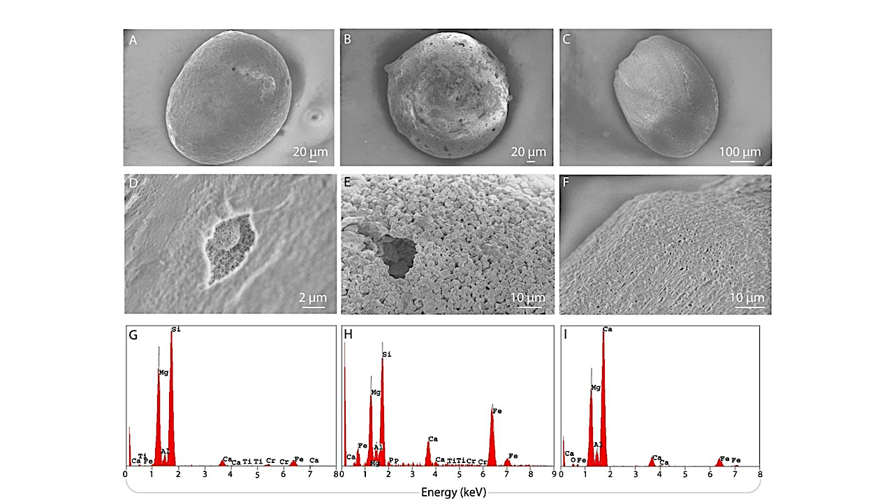

Scanning electron microscopy (SEM) and energy dispersive X-ray spectroscopy (EDX) characterization of the micrometeorites. (A-C) SEM images of the micrometeorites. (D-F) close up SEM images corresponding to (A-C). A: Porphyritic (PO), B: scoriaceous (ScMM), C: barred olivine (BO) (G-I) SEM-EDX spectra of the meteorites (intensity in counts versus energy in keV). Element assignments for each peak were provided by the instrument. Quantification of the elemental composition is presented below each spectrum. Elements which constitute more than 10% of the total are highlighted in the tables. Oxygen was not determined. — biorxiv.org
We report on the formation of membranous protocells by self-assembly of lipids on micrometeorites, the extraterrestrial particles that have been continuously reaching the surface of the Earth ever since its formation.
Synergistic interactions of lipid compartments with pristine extraterrestrial surfaces are entirely unexplored, but constitute a possible scenario for early evolution of primitive cells by a surface energy-driven transformation mechanism.
Lipids utilize the surface energy of the particles to adhere to them and autonomously transform into spherical compartments, typically through formation of lipid nanotubes. Natural sand particles of similar composition and shape were simultaneously investigated for reference, showing that certain lipid compositions prefer micrometeorite surfaces.
The elemental composition of the particles, their surface texture and cleanness altogether may be contributing to the differences observed in lipid behavior. Lipid nanotubes on- and extending out of- the micrometeorites were observed to carry lipid particles and connect to other objects in the surrounding environment.

Protocell formation on micrometeorites – Schematic description of the experimental design, including the initial step of micrometeorite collection from urban environments. Debris from roof tops containing micrometeorites is collected, and all magnetically responsive particles are separated with the help of a permanent magnet. These magnetic particles can contain micrometeorites, but other natural and man-made particulates dominate the roof top dust. Sieves are used to eliminate large particles in the millimeter range. The remaining small particles are examined under a stereo microscope and particles carrying the unique characteristics of micrometeorites are collected. The micrometeorites are then transferred into an open-top sample chamber containing aqueous medium, followed by the addition of lipid reservoirs/agglomerates. The sample becomes ready for fluorescence microscopy observation after allowing the interaction of lipids with micrometeorite surface overnight. — biorxiv.org

Protocell formation mechanism. (A-D) Formation of nanotube-lipid compartment networks on flat surfaces in aqueous environments. (A) Multilamellar lipid reservoirs, i.e., multilamellar vesicles (MLV) spread on high energy surfaces as a double lipid bilayer in the form of a flat giant unilamellar vesicle. (B) The distal lipid membrane (upper with respect to the surface) ruptures, (C) forming a lipid nanotube network. The inset to C shows the cross section of a lipid nanotube in the network. (D) Lipid compartments emerge from the lipid nanotubes. (E-I) Fluorescence microscopy images of protocells growing on model micrometeorites (plasma-treated sand particles) and micrometeorites. (E) Fluorescence micrograph of an archaeal lipid patch on a micrometeorite. The short yellow arrow points to the multilamellar reservoir. (F) Fluorescence intensity graph along the arrow in (E) (gray value from 0 to 255 of 8-bit image, distance in unit of pixels). The recorded 1:2 intensity ratio indicates single:double lipid bilayer membranes on the surface. (G) shows the vicinity of the region in (E) at three slightly different focal planes, where several lipid compartments grow out of the lipid nanotubes. The framed regions in (E) and (G) are identical. The same lipid compartment in different focal planes is marked with a yellow asterisk in (G). The nanotubes directly connected to the lipid compartments are shown with orange arrows. (H-I) fluorescence micrographs of bacteriaderived (H) and reference (I) lipids on a model micrometeorite along with inverted images for improved contrast. Magnified regions densely populated with lipid nanotubes are shown in yellow frames. The regions in orange color underscore the parts of the curved particle surface which are in focus. Blue squares attached to the images show schematically the approximate position of the protocells on the corresponding particle. — biorxiv.org
Protocell formation on micrometeorites, biorxiv.org
Astrobiology


Related Posts
Stay Informed With the Latest & Most Important News
Previous Post
Next Post


Advertisement
-
 01Two Black Holes Observed Circling Each Other for the First Time
 -
 02From Polymerization-Enabled Folding and Assembly to Chemical Evolution: Key Processes for Emergence of Functional Polymers in the Origin of Life
 -
 03Astronomy 101: From the Sun and Moon to Wormholes and Warp Drive, Key Theories, Discoveries, and Facts about the Universe (The Adams 101 Series)
 -
 04True Anomaly hires former York Space executive as chief operating officer
 -
 05Φsat-2 begins science phase for AI Earth images
 -
 06Hurricane forecasters are losing 3 key satellites ahead of peak storm season − a meteorologist explains why it matters
 -
 07Binary star systems are complex astronomical objects − a new AI approach could pin down their properties quickly